In recent years, modern medical and health-diagnostic techniques have opened the doors for stimulating developments in zoological and evolutionary studies of mollusks. In the last few decades, sophisticated genetics and molecular methodologies have been making huge contributions to the study of the “genealogy” of mollusks, or molluscan evolution. But recent adaptations and applications of health-science technologies to morphological analyses in zoology are now beginning to match the fast rate at which those molecular techniques advanced. Enter, among others, digitally reconstructed radiographs (DRR), computed tomography (CT) scans and magnetic resonance imaging (MRI). Those now familiar techniques in health diagnostics and medicine allow for quick, non-destructive and non-invasive, three-dimensional imaging, and precise three-dimensional visualizations of body parts. Now, at the close of the second decade of the second millenium, equivalent technologies such as micro-computed tomography (MCT) and micro-MRI are within the reach of zoologists and evolutionary biologists.
In a recent paper published in the American Malacological Bulletin*, Alexander Ziegler of the University of Bonn, Germany, and collaborators offer a broad sampling of images of mollusks using those and other cutting-edge techniques. The stunning images in their paper are two-dimension examples of how, using dedicated software and special digital techniques, the innards of mollusks can be viewed in three dimensions. With proper equipment, users can magnify, rotate, and tilt the animals for studies of organs and structures and how these relate to each other, all without dissecting the animal.
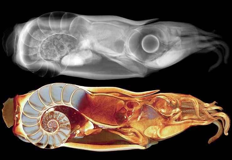
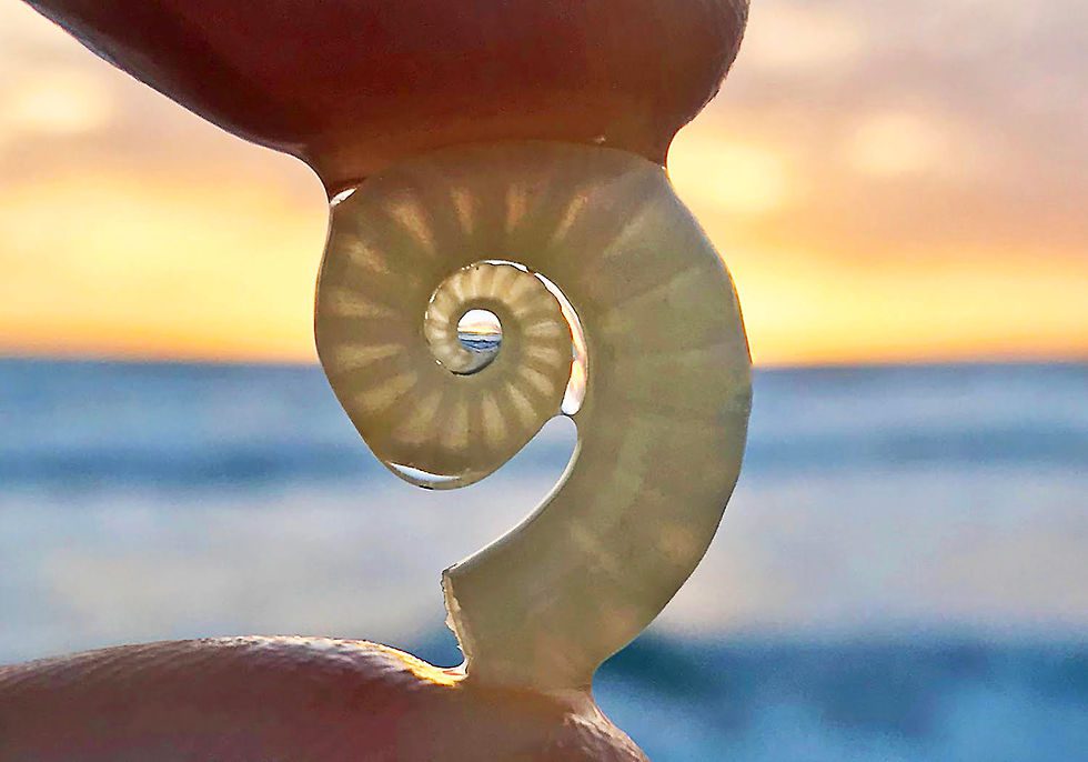
One of the species examined by Ziegler and his collaborators was the Ram Horn Squid, Spirula spirula, the 2.5-inch long, pelagic (open-water), squid-like cephalopod that makes a delicate, planispiral (“flat-coiled”) chambered shell. The images on top of the page, from Ziegler et al.’s paper, show, on top, a digitally reconstructed radiograph (DRR) and, on bottom, a micro-computed tomography rendering of a preserved Ram Horn Squid. The latter represents a “virtual slicing” of the animal; a large number of these micro-CT sections, all parallel to each other, allow for the three-dimensional reconstruction of the entire animal. (With thanks to Alexander Zeigler and collaborators for use of their data and images; more images from their study are available at the project folder in Morphobank. The photo of the Ram Horn Squid shell is from Ronald Lusk’s Instagram, and is here used with his permission.)
*Ziegler, A, C Bock, DR Ketten, RW Mair, S Mueller, N Nagelmann, ED Pracht, and L Schröder. 2018. Digital three-dimensional imaging techniques provide new analytical pathways for malacological research. American Malacological Bulletin 36(2): 248–273, 31 December 2018.